Translations:Insulin/73/ja: Difference between revisions

From Azupedia
Jump to navigation Jump to search
Created page with "=== 構造解析と合成 === {{multiple image | align = right | direction = vertical | width = 200 | image1 = Insulin monomer 4INS.png | alt1 = ブタのインスリンモノマーの白黒リボン図。 | caption1 = リチャードソン図ブタインスリンモノマーの特徴的な二次構造を示している。これがインスリンの生物学的に活性な形である。 | image2 = In..."
 
(No difference)

Latest revision as of 09:42, 19 March 2024

Information about message (contribute)
This message has no documentation. If you know where or how this message is used, you can help other translators by adding documentation to this message.
Message definition (Insulin)
=== Structural analysis and synthesis ===
{{multiple image
 | align = right
 | direction = vertical
 | width = 200
 | image1 = Insulin monomer 4INS.png
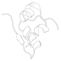
 | alt1 = Black-and-white ribbon diagram of a pig insulin monomer.
 | caption1 = [[Ribbon diagram|Richardson diagram]] of a [[pig|porcine]] insulin monomer, showing its characteristic [[secondary structure]]. This is the biologically active form of insulin.
 | image2 = Insulin hexamer 4INS.png
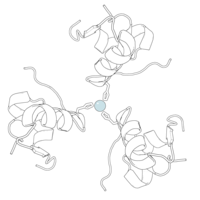
 | alt2 = Black-and-white ribbon diagram of a pig insulin hexamer, showing its characteristic quaternary structure. At the center is a pale blue-gray sphere representing a zinc atom.
 | caption2 = Richardson diagram of a porcine insulin hexamer. The sphere at the center is a stabilizing [[zinc]] atom, surrounded by coordinating [[histidine]] residues. This is the form in which insulin is stored in beta cells. {{PDB|4INS}}.
}}
Purified animal-sourced insulin was initially the only type of insulin available for experiments and diabetics. [[John Jacob Abel]] was the first to produce the crystallised form in 1926. Evidence of the protein nature was first given by [[Michael Somogyi]], [[Edward Adelbert Doisy|Edward A. Doisy]], and [[Philip Anderson Shaffer|Philip A. Shaffer]] in 1924. It was fully proven when Hans Jensen and Earl A. Evans Jr. isolated the amino acids phenylalanine and proline in 1935.

構造解析と合成

ブタのインスリンモノマーの白黒リボン図。
リチャードソン図ブタインスリンモノマーの特徴的な二次構造を示している。これがインスリンの生物学的に活性な形である。
ブタのインスリン6量体の黒と白のリボン図で、その特徴的な4次構造を示す。中心には亜鉛原子を表す薄い青灰色の球がある。
ブタインスリン6量体のリチャードソン図。中央の球は安定化亜鉛原子で、その周りをヒスチジン残基が取り囲んでいる。これはインスリンがβ細胞に貯蔵される形である。 PDB: 4INS​.

精製された動物由来のインスリンは、当初、実験や糖尿病患者に利用可能な唯一のタイプのインスリンであった。1926年にジョン・ジェイコブ・アベルが結晶化したものを初めて製造した。タンパク質の性質を示す証拠は、1924年にMichael SomogyiEdward A. DoisyPhilip A. Shafferによって初めて示された。1935年にハンス・ジェンセンとアール・A・エバンス・ジュニアがアミノ酸のフェニルアラニンとプロリンを単離したときに完全に証明された。