File:Leaf epidermis.jpg

Size of this preview: 592 × 599 pixels. Other resolutions: 237 × 240 pixels | 474 × 480 pixels | 759 × 768 pixels | 1,012 × 1,024 pixels | 2,048 × 2,073 pixels.
Original file (2,048 × 2,073 pixels, file size: 2.9 MB, MIME type: image/jpeg)
|
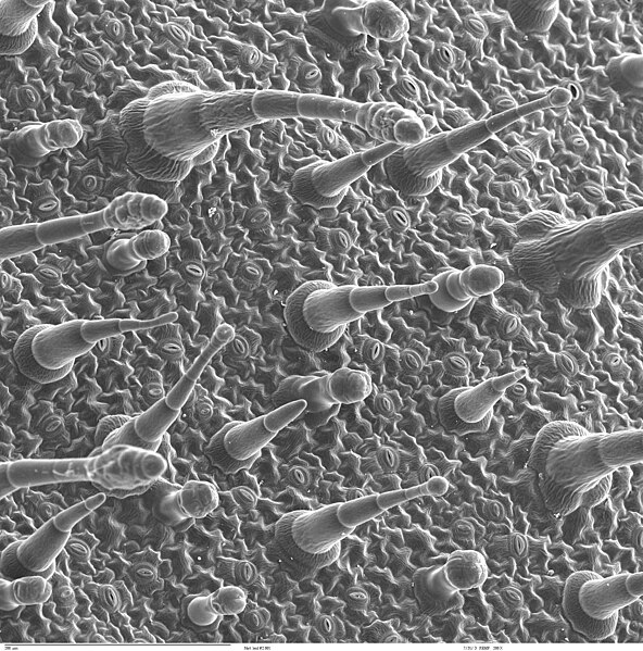
This image was selected as picture of the day on Wikimedia Commons for 14 September 2008. It was captioned as follows: English: Scanning electron microscope image of Nicotiana alata upper leaf surface, showing tricomes and a few stomates. Instrument: ZEISS962 SEM. Other languages:
Čeština: Snímek povrchu listu rostliny Nicotiana alata pořízený rastrovacím elektronovým mikroskopem, jsou zde vidět např. trichomy a průduchy. Přístroj: ZEISS962 SEM. Dansk: Billede af bladoverside, taget af elektronmikroskop og visende trichomer og nogle få stomata. Deutsch: Ansicht der Blattoberfläche des Nachtschattengewächses Nicotiana alata durch ein Elektronenmikroskop ZEISS962 SEM. Hier sichtbar: einige Trichome und Stomata English: Scanning electron microscope image of Nicotiana alata upper leaf surface, showing tricomes and a few stomates. Instrument: ZEISS962 SEM. Español: Imagen de la superficie de una hoja de tabaco (Nicotiana alata) por un microscopio electrónico de barrido, mostrando algunos tricomas y estomas. Instrumento: ZEISS962 MEV. Esperanto: bildo pri la surfaco de tabaka folio, vidataj per elektrona mikroskopo - videblas kelkaj trihxomoj (plantoharoj) kaj stomoj Français : Image par microscopie électronique de la surface supérieure des feuilles de Nicotiana alata, montrant des trichomes et quelques stomates. Instrument : ZEISS962 SEM Italiano: Immagine al microscopio elettronico a scansione della pagina superiore di una foglia di tabacco (Nicotiana alata). Magyar: A díszdohány (Nicotiana alata) levéllemezének pásztázó elektronmikroszkóppal készült képe, amelyen jól kivehetőek a szőrképletek (trichómák) és a szem alakú gázcserenyílások (sztómák) Nederlands: Aanzicht van het bladoppervlak van siertabak door het oog van een elektronenmicroscoop van het type ZEISS962 SEM, waarbij haren en huidmondjes zichtbaar zijn Polski: Obraz górnej powierzchni liścia tytoniu oskrzydlonego (Nicotiana alata) z widocznymi włoskami i szparkami, uzyskany przy pomocy elektronowego mikroskopu skaningowego ZEISS962 SEM. Português: Imagem da superfície de uma folha de tabaco (Nicotiana alata) por um microscópio eletrônico de varredura, mostrando tricomas e alguns estômatos. Instrumento: ZEISS962 MEV. Svenska: Ovansidan av ett blad av blomstertobak (Nicotiana alata), fotograferat med ett svepelektronmikroskop. Русский: Лист табака крылатого (Nicotiana alata) под электронным микроскопом ZEISS962 SEM. Видны трихомы и устьица Українська: Вигляд листя виду тютюну (Nicotiana alata тютюн крилатий) під електронним мікроскопом марки ZEISS962 SEM. 한국어: Nicotiana alata의 위쪽 잎을 현미경으로 관찰한 모습을 스캔한 것. 돌기와 기공이 보인다. 기구: ZEISS962 SEM. 日本語: ニコチアナ(Nicotiana alata)の葉の表側の電子顕微鏡写真 中文: 电子显微镜下烟草叶子的表面,可以看到气孔。 |
| Description | Scanning electron microscope image of Nicotiana alata upper leaf surface, showing tricomes and a few stomates. Instrument: ZEISS962 SEM. | ||
| Date | Unknown date | ||
| Source | http://remf.dartmouth.edu/images/NicotianaLeafSEM/nicotianaleafcatalog.html | ||
| Author | Louisa Howard (Dartmouth electron microscope facility) | ||
| Permission (Reusing this file) |
|
||
| Other versions | Derivative works of this file: Leaf epidermis w scale.jpg |
File history
Click on a date/time to view the file as it appeared at that time.
| Date/Time | Thumbnail | Dimensions | User | Comment | |
|---|---|---|---|---|---|
| current | 03:26, 22 June 2008 | | 2,048 × 2,073 (2.9 MB) | wikimediacommons>Calliopejen1 | {{Information |Description=Scanning electron microscope image of Nicotiana alata upper leaf surface, showing tricomes and a few stomates. Instrument: ZEISS962 SEM. |Source=http://remf.dartmouth.edu/images/NicotianaLeafSEM/nicotianaleafcatalog.html |Date=U |
File usage
There are no pages that use this file.